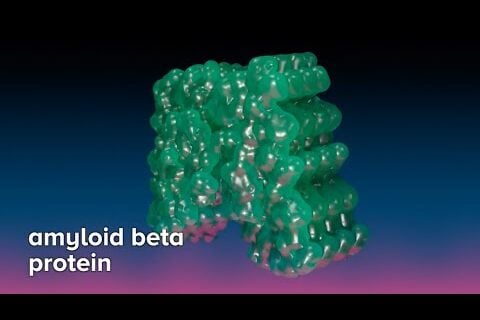
Alzheimer’s plaque is caused by proteins that aggregate together. Protein aggregation is a hallmark of many neurodegenerative diseases such as Alzheimer’s. However, the mechanisms linking aggregation to Alzheimer’s neurotoxicity remain poorly understood.
One reason is because only limited information is available on the structure of these aggregates. Determining the atomic structures of these aggregates is crucial to understanding their formation, clearance and spread in the human brain. It makes sense that this even holds the important key to unlocking better Alzheimer’s treatments. (Article continued below…)
Furthermore, very little is known about the native structure of protein aggregates inside cells. This challenge is addressed utilizing the latest developments in world-class microscopes using cryo-electron tomography (cryo-ET). Thin lamellas of vitrified cells containing protein aggregates are prepared by cryo-focused ion beam (cryo-FIB) and subsequently imaged in three dimensions by cryo-ET.
Cryo-Electron Microscopy is helping Dr. Anthony Fitzpatrick’s lab at Columbia University in New York to untangle amyloid fibrils isolated from post-mortem brain tissue of patients with Alzheimer’s and other types of dementia.
These super-high-powered microscopes elucidate the molecular and structural basis of neurodegeneration. Additionally, cryo-ET allows for the analysis of aggregate structures within pristinely preserved cellular environments at molecular resolution, shedding new light on the cellular mechanisms of neurodegeneration.


