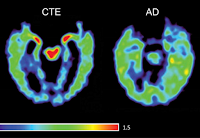
DIAGNOSIS NEWS: CTE dementia can now be diagnosed with certainty. (CTE [Chronic Traumatic Encephalopathy] is caused by blows to the head.) This differs from the clinical diagnosis for dementias such as Alzheimer’s, which are based on a doctor’s best judgement call of tests and interviews. Learn how doctors can be sure when it’s CTE.
The main characteristic of CTE is the abundance of protein “Tau tangles.” Large numbers of tau tangles also exist in Alzheimer’s disease. Historically, it was believed that CTE and Alzheimer’s tau tangles were made in the same way. Instead, what an international research team found (and reported March 20 in the peer-reviewed scientific journal Nature) is a much different story.
How to Tell It’s CTE
The main characteristic of CTE is the abundance of the aggregated insoluble tau protein, which results in the formation of tau tangles in an individual’s brain cells.
Using cryogenic electron microscopy (cryo-EM), the researchers examined tau tangles extracted from the brains of an American football player and two professional boxers, all recognized neuropathologically as having suffered from CTE.
Different Folds for Different Dementias
What they determined is that the fold of the abnormal tau protein in CTE is different from the tau fold in Alzheimer’s disease. This same research team discovered the Alzheimer’s disease tau fold in 2017, a discovery which was featured on the cover of Nature.
Bernardino Ghetti, MD, neuropathologist and member of the research team, noted the importance of the finding: “The use of Cryo-EM has allowed us to determine the 3D structure of tau and facilitated our understanding of how it functions and interacts. Understanding these differences will accelerate the discovery of substances that may prevent the formation of abnormal tau in the brain cells of people suffering from either of these two distinct neurodegenerative diseases.”
Two New CTE Discoveries
Additionally, the research team discovered an unidentified element adjacent to the tau of CTE, which does not exist in the Alzheimer’s tau.
Ruben Vidal, Ph.D., professor in the Department of Pathology and Laboratory Medicine at IU School of Medicine, said, “These two new discoveries provide more insights into CTE than had previously existed. The information will be incredibly valuable for the development of novel agents to help in diagnosis and therapeutics specifically designed for individuals fighting CTE.”
Consensus Panel Agrees on CTE
In further research, a consensus panel of expert neuropathologists confirmed CTE as a unique disease that can be definitively diagnosed by neuropathological examination of brain tissue. They concluded that CTE has a pathognomonic signature in the brain. This is an advance that represents a milestone for CTE research and lays the foundation for future studies defining the clinical symptoms, genetic risk factors and therapeutic strategies for CTE.
The neuropathological criteria defining CTE, or the NINDS CTE criteria, which appear in the journal Acta Neuropathologica, was defined at the Foundation of the National Institutes of Health (NIH).
Head Hits
CTE is a progressive degenerative disease of the brain found in persons with a history of repetitive brain trauma, including symptomatic concussions as well as asymptomatic sub-concussive hits to the head. The trauma triggers progressive degeneration of the brain tissue, including the build-up of an abnormal protein called tau. These changes in the brain can begin months, years or even decades after the last brain trauma or end of active athletic involvement. The brain degeneration is associated with memory loss, confusion, impaired judgment, impulse control problems, aggression, depression, and, eventually, progressive dementia.
CTE has a Unique Signature
The consensus panel of seven neuropathologists independently reviewed slides from 25 cases of different diseases associated with tau deposits in the brain, completely blinded to all clinical information, including age, sex, clinical symptoms and athletic exposure using provisional diagnostic criteria for CTE developed by Ann McKee, MD, Director of the CTE Program at Boston University and Chief of Neuropathology. The neuropathologists concluded that the criteria distinguished CTE from other tauopathies, including aging and Alzheimer’s, and that CTE had a unique pathological signature in the brain.
According to McKee, neuropathologists agreed on the diagnosis of CTE and confirmed the interim standards. “The specific feature considered unique to CTE was the abnormal perivascular accumulation of tau in neurons, astrocytes and cell processes in an irregular pattern at the depths of the cortical sulci,” explained McKee who is corresponding author of the study. “This lesion was not characteristic of any of the other disorders, including Alzheimer’s disease, age-related tauopathy or progressive supranuclear palsy, and has only been found in individuals who were exposed to brain trauma, typically multiple episodes,” she added.
- The international team reporting these findings includes, Michel Goedert, MD, Ph.D., Sjors Scheres, Ph.D., Benjamin Falcon, PhD and their collaborators in the Laboratory of Molecular Biology of the Medical Research Council (Cambridge, UK), plus Bernardino Ghetti, MD, Ruben Vidal, Ph.D., and Holly Garringer, Ph.D. at the Dementia Laboratory at the Department of Pathology and Laboratory Medicine at IU School of Medicine, and Kathy Newell, MD, currently at the University of Kansas, and who will join the Department of Pathology and Laboratory Medicine at IU School of Medicine in June.
SUPPORT:
- Research conducted in the United States was funded by the U.S. National Institutes of Health, IU School of Medicine and the University of Kansas School of Medicine. Research conducted in the United Kingdom was funded by the U.K. Medical Research Council (MRC), and the European Union.
SOURCES:
REFERENCES:
- Benjamin Falcon, Jasenko Zivanov, Wenjuan Zhang, Alexey G. Murzin, Holly J. Garringer, Ruben Vidal, R. Anthony Crowther, Kathy L. Newell, Bernardino Ghetti, Michel Goedert, Sjors H. W. Scheres. Novel tau filament fold in chronic traumatic encephalopathy encloses hydrophobic molecules. Nature, 2019; DOI: 10.1038/s41586-019-1026-5
- Ann C. McKee, Nigel J. Cairns, Dennis W. Dickson, Rebecca D. Folkerth, C. Dirk Keene, Irene Litvan, Daniel P. Perl, Thor D. Stein, Jean-Paul Vonsattel, William Stewart, Yorghos Tripodis, John F. Crary, Kevin F. Bieniek, Kristen Dams-O’Connor, Victor E. Alvarez, Wayne A. Gordon.The first NINDS/NIBIB consensus meeting to define neuropathological criteria for the diagnosis of chronic traumatic encephalopathy. Acta Neuropathologica, 2015; DOI: 10.1007/s00401-015-1515-z


As is well known,the main neurons disorders that leads to neurodegeneration in Repeated Concussion and in Chronic Traumatic Encephalopathy,are the biochemical,molecular, metabolical and brain energy disorders,that leads to tau hyperfosforilation and tau accumulation.As we can read in the reference articles bellow ,in animal models, doses of only fifty miligrams by day/rat,per os,of ACETYL L CARNITINE(ALCAR),a constituent of the inner mitochondrial membrane, enhances memory retention,LOWERS TAU PHOSPHORYLATION AND ACCUMULATION(1),(2),(3)and LOWERS BETAMYLOID ACCUMULATION(3).ALCAR looks to fits for research as support therapy (and maybe to prevent?)the taupathies related to repeated concussion and to the Chronic Traumatic Encephalopathy.
1) "ACETYL-L-CARNITINE ATTENUATES OKADAIC ACID INDUCED TAU HYPERPHOSPHORYLATION AND SPATIAL MEMORY IMPAIRMENT IN RATS – Journal of Alzheimer Disease, in 2010,author Yin YY and colleagues 2) "ACETYL-L-CARNITINE AMELIORATES SPATIAL MEMORY DEFICITS INDUCED BY INHIBITION OF PHOSPHOINOSITOL-3 KINASE AND PROTEIN KINASE C" , Journal of Neurochemistry, in 2011 ,Jiang X and colleagues 3) "ACETYL-L-CARNITINE ATTENUATES HOMOCYSTEINE-INDUCED ALZHEIMER-LIKE HISTOPATHOLOGICAL AND BEHAVIORAL ABNORMALITIES" Zhou P and colleagues